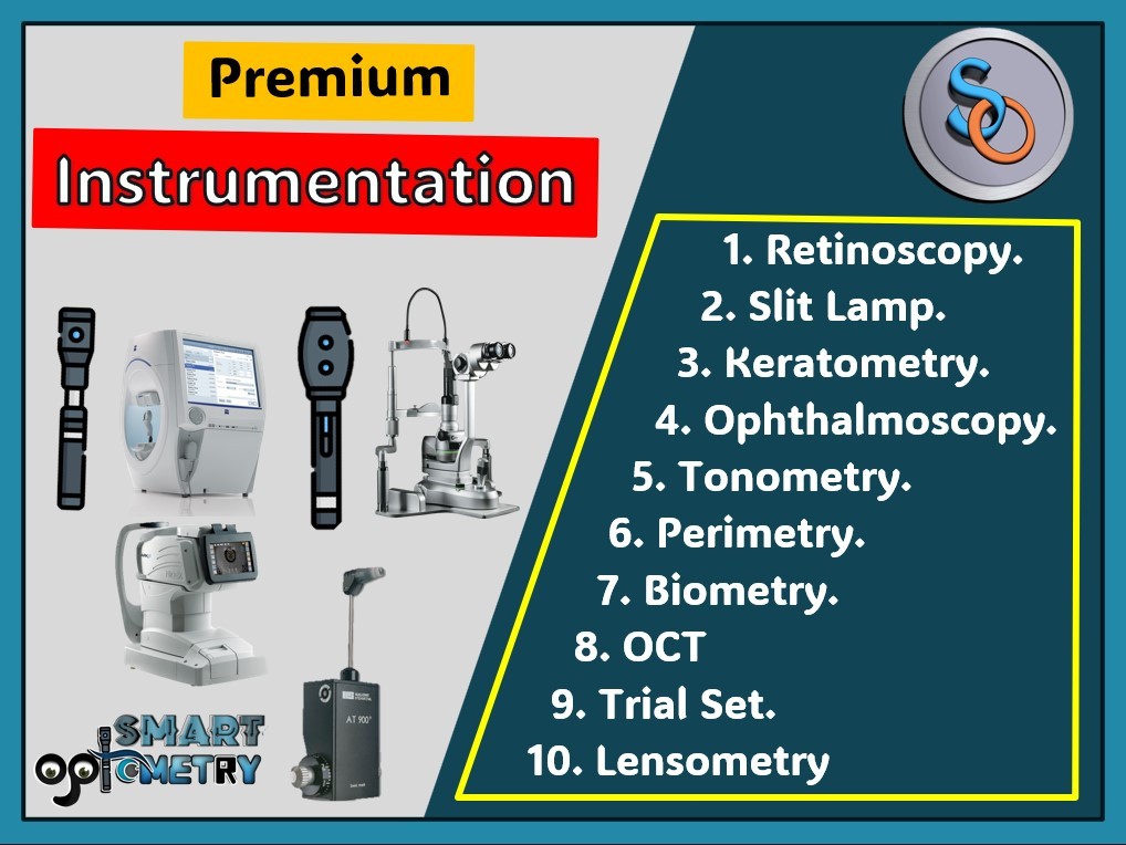
1. Retinoscopy: PDF-1. Overview of retinoscopy: This PDF will cover: • What is Retinoscopy. • Principle of Retinoscopy. • Types of Retinoscope. • Spot vs Streak Retinoscope • Parts of Streak Retinoscope • Characteristics of RET reflex. PDF-2: Working Distance & Working Distance power: • What is working distance in retinoscopy. • What is working distance power in retinoscopy? • Why do we need to subtract working distance power? • How to identify working distance power at different distances. PDF-3: Methods of Streak Retinoscopy. • Method-1: Retinoscopy with 1 spherical & 1 Cylinder Trail lens. • Method-2: Retinoscopy with 2-spherical trial lenses. • Method-3: Retinoscopy with 2-cylinder trial lenses. PDF-4: Cross Retinoscopy. • This PDF will cover how to do Cross retinoscopy (retinoscopy with 2 spherical lense) just in 4 simple steps with example. PDF-5: Cycloplegic refraction: This PDF will cover details about: • Why Cycloplegic Refraction? • Principle and Mechanism of Cycloplegic Refraction. • Properties of Cycloplegics Drugs. • Indications Cycloplegic refraction. • Contraindications Cycloplegic refraction. • Side Effects Cycloplegic drugs/refraction. • Procedure of Cycloplegic refraction. • Post Mydriatic Treatment. • How to Choose ideal Cycloplegics. • Spectacle Prescribing in cycloplegic refraction. Videos: • Optical Cross in Retinoscopy. • How to calculate Retinoscopic power. • How to practice retinoscopy with simulator. 2. Direct Ophthalmoscopy- This PDF will cover: • Why do we not able to see patient’s retina with naked eye? • Definition & Principle of Direct Ophthalmoscopy. • Structure of Direct Ophthalmoscope. • Aperture available in Direct Ophthalmoscope. • Indications of Direct Ophthalmoscope. • Procedure of Direct Ophthalmoscope. 3. Biometry: PDF-1: Biometry- An Overview. • Introduction of Biometry. • Indications of Biometry. • Ultrasonic Method: Contact & Immersion. • Optical Biometry in Brief. • Effective IOL position. • How to calculate IOL power. • Error in IOL calculation. PDF-2: Biometry Formulas. • Introduction of Biometry Formula. • Historical/Refraction based Formulas. • Theoretical Formulas. • Regression Formulas. • Vergence Formulas. • AI Formulas. • Ray tracing Formulas. PDF-3: Generations of Biometry Formulas. • First-Generation Formulae. • Second-generation Formulae. • Third Generation Formulae. • Fourth-generation Formulae. • Fifth-generation Formulae. • Newer IOL Power Calculation Formulae. • Barrett Universal Formula. • Olsen Formula. • Hill-RBF Formula. 4. Perimetry: PDF-1: Perimetry- An Overview. • What is Visual Field. • Types of Visual Field Defect. • What is Perimetry. • Indications of Perimetry. • Kinetic Vs Static Perimetry. • Central Vs Peripheral Field Charting. • Manual Vs Automated Perimetry. • Procedure of Manual Perimeter. • Procedure of Automated Perimeter. • How does Software detect Visual Field (VF) defects. PDF-2: Interpret Humphrey Visual Field Report. • Patient data and test parameters. • Reliability indices. • Grey Scale. • Total deviation plots. • Pattern deviation plots: • Global indices. • Glaucoma hemifield test (GHT). • Actual threshold values. • Does Clinician follow all these parameters to evaluate visual Field Reports? 5. Slit-Lamp Biomicroscope. PDF-1: Slit-Lamp- An overview. • Introduction of Slit Lamp. • Observable Parts of Slit Lamp. • Types of Slit Lamp. • Principle of Slit Lamp. • Observation System of Slit Lamp. • Illumination System of Slit Lamp. • Mechanical System of Slit Lamp. PDF-2: Procedure & illumination system of Slit Lamp. • Procedure of Slit Lamp. • Diffuse illumination. • Direct illumination. • Optical section • Parallelopiped • Conical beam • Indirect illumination. • Retro-illumination • Specular reflection. • Sclerotic Scatter. • Oscillating illumination. 6. Tonometry. PDF-1: Tonometry - An Overview. • Introduction to tonometry. • Types of Tonometry. • Direct Vs Indirect Tonometry. • Contact Vs Non-Contact Tonometry. • Applanation Tonometry. • Indentation Tonometry. • Palpation Tonometry. • Indication of Tonometry. • Contraindication of Tonometry. PDF-2: Goldmann Applanation Tonometer (GAT). • Introduction of Goldmann Applanation Tonometer. • Principle of Goldmann Applanation Tonometer. • Parts of Goldmann Applanation Tonometer. • Procedure of Goldmann Applanation Tonometer. • Interpretation of Goldmann Applanation Tonometer. • Source of error of Goldmann Applanation Tonometer. 7. Trial Set- A Complete Tutorial: • Introduction of trial frame. • Trial Lenses: Full Aperture & Reduced Aperture. • Trial Frame: Full, Reduced & Half Eye Frame. • Prism: Range & usage. • Plano Lens & Occluder. • Pin Hole: Why does vision improve with Pin Hole. • Maddox Rod: Red & White Maddox rod. • Near Vision Chart: Snellen near vision chart. • Stenopaeic Slit: Identifying Astigmatism, • Emsley Fincham test • Red-Green Filter: Worth Four Dot Test. • Jackson Cross C